top of page
Ara


Acute Idiopathic Maculopathy
An acute inflammatory disease of the retina pigment epithelium


Relentless Placoid Chorioretinitis
White dot syndrome


Central Areolar Choroidal Dystrophy (CACD)
Hereditary disease of the choroid

Combined choroidal neovascularization
Combined type 1 and type 2 choroidal neovascularisation in armd


Sympathetic ophthalmia
Posttraumatic uveitis


BALAD
Bacillary layer detachment, extensive changes in structure of the retina

Retinal Angiomatous Proliferation (RAP)
neovascularisation in the retina, armd


Peripapillary Choroidal Neovascularisation
Choroidal neovascularisation originating from the peripapillary region


Subfoveal Choroidal Neovascular Membrane
Armd with choroidal neovascularisation underneath the fovea.


Juxtafoveal Chorodial Neovascular Membrane
Choroidal neovascularisation adjacent to foveal region


Type 1 Neovascularisation
Most frequently encountered choroidal neovascularisation type in armd


Idiopathic Polypoidal Choroidal Vasculopathy
A rare subtype of armd, there are more cases diagnosed in recent years.

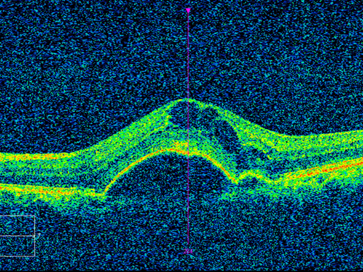
Vascularized Pigment Epithelial Detachment
May be accepted as precursor findings of choroidal neovascularisation

Pigment Epithelial Detachment
Mostly encountered in armd but other etiologies must be considered in differential diagnosis

Retinal Pigment Epithelial Tears
A rare but devastating complication of both the disease itself and treatment in armd.
bottom of page
